atlas and epitome

*
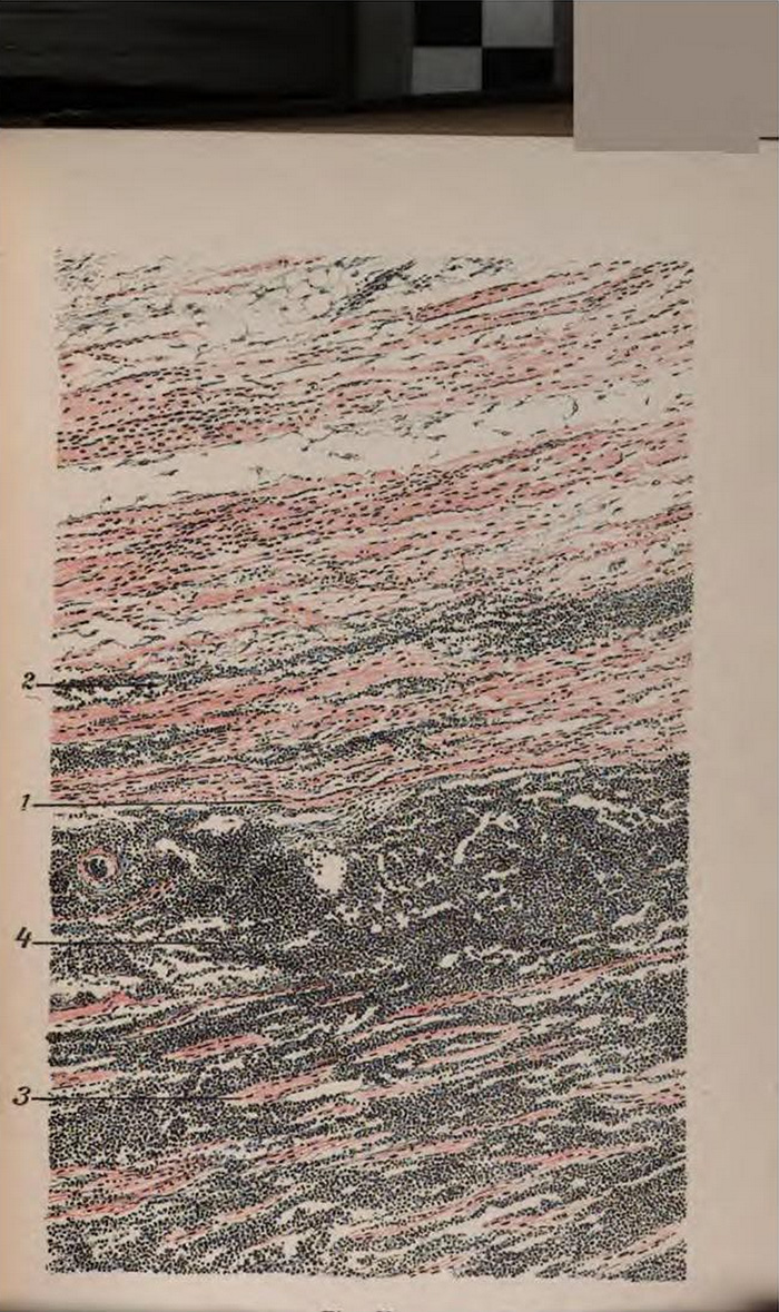
Figure X.
Infiltrating Growth of a Small-cell Sarcoma in Muscle (x 70.)
in Hermann Dürck. Atlas and Epitome of General Pathologic Histology. Authorized translation from the German. Edited by Ludvig Hektoen. (1904)
Stanford (Lane Medical Library) copy, archive.org scan, no date of digitization
Shown above is the other view mentioned in my asfaltics post, that gave rise to the ancient name
The three volumes of Dürck’s Atlas and Epitome are remarkable. Here are links to all three:
-
Hermann Dürck. Atlas and Epitome of Special Pathologic Histology.
Circulatory Organs; Respiratory Organs; Gastro-Intestinal Tract
With 62 Colored Plates
Authorized translation from the German. Edited by Ludvig Hektoen. (1900)
Stanford (Lane Medical Library) copy
Google Books
archive.org
Library of Congress copy
archive.org - Hermann Dürck. Atlas and Epitome of Special Pathologic Histology.
Liver; Urinary Organs; Sexual Organs; Nervous System; Skin; Muscles; Bones
With 123 Colored Illustrations on 60 Lithographic Plates
Authorized translation from the German. Edited by Ludvig Hektoen. (1901)
Stanford (Lane Medical Library) copy, no date of digitization
Google Books
archive.org
Harvard copy
archive.org - Hermann Dürck. Atlas and Epitome of General Pathologic Histology.
With 176 Colored Illustrations on 80 Lithographic Plates and 36 Figures in Black and Colors.
Authorized translation from the German. Edited by Ludvig Hektoen. (1904)
Stanford (Lane Medical Library) copy
Google Books
archive.org
Wikipedia pages on
Hermann Dürck (1869-1941) and
Ludvig Hektoen (1863-1951)
tags:
dots; lines; seas; waves; H. Dürck, Atlas and Epitome of General Pathologic Histology (1904); L. Hektoun